What OpenDerm is
OpenDerm is an open-source robotic imaging system for creating high-resolution, reproducible 3D maps of the skin. It was designed and built by Marion Lepert, and the hardware designs, software, and build documentation are all openly available.
"Reproducible" is the load-bearing word. Comparing registered scans — scans aligned to the same positions — is meant to reveal new lesions and identify subtle changes in existing ones. The design is built around imaging the same skin repeatedly under the same conditions, not around a single capture.
The project also states plainly that this is not a medical device and is for research use only. It is not positioned as a diagnostic tool.
OpenDerm is an open-source robotic imaging system for creating high-resolution, reproducible 3D maps of the skin. Registered scans help reveal new lesions and identify subtle changes in existing ones. The hardware designs, software, and build documentation are all openly available. / Designed and built by Marion Lepert. / Not a medical device · Research use only — From the statements on what the project is and what it publishes, on who built it, and on the limits of its intended use
High resolution and low cost at the same time
The numbers make the positioning clear. Capture resolution is 78 pixels per millimeter, enough to resolve details such as a mole's pigment network and the individual hairs next to it.
Hardware comes to less than $8,500. The project states that this provides some of the highest-resolution imaging available at a fraction of the cost of commercial total-body photography systems. Holding both of those at once is the claim.
OpenDerm captures skin at 78 pixels per millimeter, resolving details such as a mole’s pigment network and nearby individual hairs. / OpenDerm captures skin at 78 pixels per millimeter using less than $8,500 in hardware. It is fully open source and provides some of the highest-resolution imaging available at a fraction of the cost of commercial total-body photography systems. — From the statements on positioning, capture resolution, and cost
Two licenses, split by what they cover
Being buildable is part of the design. What is published is the complete bill of materials, the CAD assembly, electrical and safety schematics, and step-by-step build instructions.
The licenses split by target. Software is under the MIT License. Hardware design files, schematics, bill of materials, and build documentation are under CERN-OHL-P-2.0 (CERN Open Hardware Licence Version 2 - Permissive), an open-hardware license whose Permissive variant is the one chosen here.
The complete bill of materials, CAD assembly, electrical and safety schematics, and step-by-step build instructions are publicly available. / OpenDerm software is available under the MIT License. The hardware design files, schematics, bill of materials, and build documentation are available under the CERN Open Hardware Licence Version 2 - Permissive (CERN-OHL-P-2.0). — From the statements on what documentation is published and on the software and hardware licenses
How it is built
Looking at the construction explains how the price lands where it does.
Off-the-shelf parts on four axes
OpenDerm is built primarily from off-the-shelf parts. Keeping custom fabrication to a minimum is the design choice that holds the cost down.
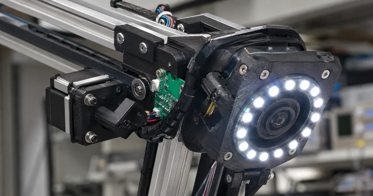
The camera rides on a gantry with four degrees of freedom. Three linear axes handle positioning: X travels along the side rails, Y moves across the top beam, and Z sets the camera height. A rotary axis, RX, tilts the sensor head to align the camera with the skin surface.
OpenDerm's four degrees of freedom (per the official site)
OpenDerm is built primarily from off-the-shelf parts. / The gantry moves the camera through four degrees of freedom. Three linear axes position the sensor head within the workspace: X travels along the side rails, Y moves across the top beam, and Z sets the camera height. A rotary axis (RX) tilts the sensor head to align the camera with the skin surface. — From the statements on the off-the-shelf construction and on the four degrees of freedom
What sits on the sensor head
The imaging assembly is documented just as concretely. Four things ride on it.
A Canon EOS R7 body with an RF 100 mm macro lens. Lighting comes from a Godox MF-R76 ring flash fitted with a cross-polarization filter, which cuts specular glare so the image shows structure below the surface rather than reflections off it.
And two downward-facing laser distance sensors. Those two are the point: their measurements let the control system maintain a consistent working distance and camera orientation relative to the skin. The goal of shooting the same conditions repeatedly leads directly to this parts list.
The capture flow is documented as well. At each imaging station the robot uses the two laser sensors to set the camera's angle and working distance, then takes a high-resolution photograph. The pipeline afterwards aligns the overlapping images and reconstructs them as one continuous surface.
The sensor head carries a Canon EOS R7 with an RF 100 mm macro lens, a Godox MF-R76 ring flash fitted with a cross-polarization filter that reduces specular glare, and two downward-facing laser distance sensors. The distance measurements allow the control system to maintain a consistent working distance and camera orientation relative to the skin. / At each imaging station, the robot uses two laser sensors to control the camera’s angle and working distance before capturing a high-resolution photograph. The complete pipeline then aligns the overlapping images and reconstructs them as one continuous surface. — From the statements on what the sensor head carries and on the capture flow
Why move a robot instead of adding cameras
One camera in motion rather than a large array of fixed ones. The reasoning is stated.
By moving close to the body and following its contours, the system can maintain consistent distance, angle, focus, and lighting while capturing high-resolution images across the skin surface. The second reason is cost: using a single camera with cheap actuators reduces hardware cost compared with a large array of fixed cameras.
A robotic imaging system offers a practical way to meet these requirements. By moving close to the body and following its contours, the system can maintain consistent distance, angle, focus, and lighting while capturing high-resolution images across the skin surface. Using a single camera with cheap actuators also reduces hardware cost compared with a large array of fixed cameras. — From the statement on why a robotic approach was chosen
What makes this an AI story
The reason this project shows up in an AI context has less to do with the imaging rig than with what kind of data it is trying to produce.
The weakness in existing training data
The project names a structural problem with AI models for skin-cancer detection.
Most of them are trained on isolated images of lesions already identified as suspicious. Those datasets therefore provide little evidence of how melanomas first emerge and evolve before drawing clinical attention.
Recognizing earlier signs would require repeated, high-resolution images of the same skin over time. That is why OpenDerm insists on reproducibility and on making registered scans comparable. Building a machine is not the goal; getting a machine that can collect that kind of data into wide, affordable circulation is.
Most AI models for skin-cancer detection are trained on isolated images of lesions already identified as suspicious. These datasets therefore provide little evidence of how melanomas first emerge and evolve before drawing clinical attention. Training models to recognize earlier signs of melanoma will require repeated, high-resolution images of the same skin over time. / Total-body photography must become more affordable and widely available. Patients at high risk should be able to receive frequent, standardized scans. — From the statements on the limits of existing datasets, on the kind of data needed, and on the accessibility argument in the Frequent, accessible scanning card
Where it sits among three approaches
The project frames wide-area skin imaging as three approaches, each trading speed, detail, consistency, footprint, and cost differently.
Three approaches to wide-area skin imaging (per the official site)
| Approach | Speed | Detail | Cost | Examples given |
|---|---|---|---|---|
| Wide-field total-body photography | Seconds | Wide-field detail | $$$ | Neko / Canfield VECTRA / DermSpectra / FotoFinder |
| Close-range robotic scanning | Minutes | High-resolution detail | $$ | SquareMind / iToBoS / OpenDerm |
| Smartphone-guided capture | Manual | Variable detail | $ | SkinIO / MoleMap / Miiskin |
OpenDerm belongs to the middle row, close-range robotic scanning. A high-resolution camera on a robotic arm or gantry moves close to the skin and systematically across the body while holding distance and viewing angle under control. Region by region, a single camera captures much finer detail than a distant wide-field system, using less space and less camera hardware. The trade-off is speed: the body has to be scanned sequentially rather than all at once.
The project also notes that the field itself is still emerging, with limited clinical availability in the United States and many systems still experimental or under development.
Total-body photography remains an emerging field, with limited clinical availability in the United States and many systems still experimental or under development. / A high-resolution camera mounted on a robotic arm or gantry moves close to the skin and systematically across the body while maintaining a controlled distance and viewing angle. Imaging the skin region by region allows a single camera to capture much finer detail than a distant wide-field system, while requiring less space and camera hardware. The trade-off is speed: the body must be scanned sequentially rather than all at once. / Seconds Wide-field detail $$$ Examples: Neko · Canfield VECTRA · DermSpectra · FotoFinder / Minutes High-resolution detail $$ Examples: SquareMind · iToBoS · OpenDerm / Manual Variable detail No dedicated hardware $ Examples: SkinIO · MoleMap · Miiskin — From the statements on the state of the field, on the characteristics and trade-off of close-range robotic scanning, and on the speed, detail, cost, and examples listed in the Approach 01–03 cards
The problem it starts from
The site opens by stating what the project takes as given. We reproduce the published figures as published and stop short of any medical interpretation.
The figures listed are that more than 100,000 Americans are diagnosed with melanoma each year; that the five-year relative survival rate is nearly 100% when detected early but about 34% after it reaches distant parts of the body; and that approximately 30% of melanomas develop within an existing mole while 70% appear as new lesions. The site cites sources for each.
From there it names three areas where progress is needed: higher resolution imaging, longitudinal datasets, and frequent, accessible scanning. OpenDerm reads as an attempt to satisfy all three at once. When you are working through technical documentation like this, converting the pages into a readable local format makes it easier to move between the figures and the text.
More than 100,000 Americans are diagnosed with melanoma each year. This form of skin cancer has a five-year relative survival rate of nearly 100% when detected early, but about 34% after it reaches distant parts of the body. … Approximately 30% of melanomas develop within an existing mole, while 70% appear as new lesions. / Improving total-body imaging requires progress in three areas: … Higher Resolution Imaging / Longitudinal datasets / Frequent, accessible scanning — From the figures the project starts from and the three areas it names as needing progress
Wrapping up: the cheap machine matters less than the data it makes possible
The eye-catching part of OpenDerm is the "under $8,500." But the significance is less that a cheap machine exists and more what it makes possible downstream.
As the project points out, most AI models for skin-cancer detection learn from isolated images of lesions that were already flagged as suspicious. The moment a lesion appears, and the changes before anything drew attention, are simply absent from the data. Recognizing an earlier stage requires repeated images of the same skin — and that data does not accumulate while the machines stay expensive and scarce.
The engineering lesson is that the constraints drove the design. Because the same conditions have to be reproduced, laser distance sensors hold distance and angle constant. Because cost has to come down, one camera moves instead of many staying still. Speed is what gets given up. What is traded for what is stated outright.
Design files, schematics, bill of materials, and instructions are all published, under MIT for software and the permissive CERN-OHL-P-2.0 for hardware. That makes it readable as a template for carrying the same approach to a different subject.
The same thread — that the scaffolding around a model does the work — runs through the system Google built to fix Chrome vulnerabilities and how context engineering is approached. For open-weight model releases, see Kimi K3 and Inkling-Small.